
Partner news: Combine 3D models and X-ray images with the latest release of Mimics®
June 20, 2014
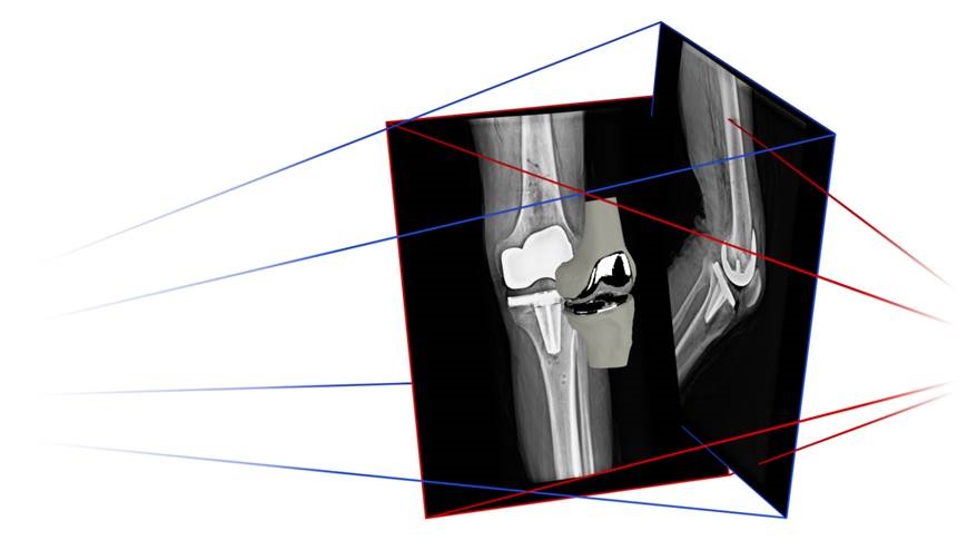
A new X-ray module has been added to the Mimics® Innovation Suite (Materialise NV) which allows its users to align X-ray images with 3D models and determine 3D position of bone and implant models based on X-ray images. The software suite which is best known for the segmentation of CT or MRI images to create 3D models and use them for analysis, for design of patient specific devices or to create finite element meshes, now opens up an entire new range of possibilities.
Being able to analyze the 3D position of bones and implants at different points in time or in different conditions without the need of multiple CT or MRI scans, but instead using commonly available X-ray images, drastically increases the amount of clinical data engineers can use in their workflow. This is for example a big leap forward for anyone who is working with 3D surgical planning techniques like patient specific guides, because besides planning and designing in 3D, they can now finally also measure the post-operative result in 3D. This allows to finally close the engineering cycle, allowing researchers to better analyze and improve their innovations.
In a recent Materialise webinar, guest speakers from the Hospital of Special Surgery in New York and from the orthopedic company Zimmer explain how they used this new module for custom implant evaluation and in the design of a shoulder implant system respectively.
For further information visit the Materialise website or email [email protected]
- July 2019
- February 2019
- December 2018
- November 2018
- October 2018
- August 2018
- July 2018
- June 2018
- May 2018
- April 2018
- March 2018
- February 2018
- January 2018
- December 2017
- November 2017
- October 2017
- September 2017
- August 2017
- June 2017
- May 2017
- March 2017
- February 2017
- January 2017
- November 2016
- October 2016
- September 2016
- July 2016
- June 2016
- May 2016
- April 2016
- March 2016
- February 2016
- January 2016
- December 2015
- November 2015
- September 2015
- July 2015
- June 2015
- May 2015
- April 2015
- March 2015
- February 2015
- January 2015
- December 2014
- November 2014
- October 2014
- September 2014
- August 2014
- July 2014
- June 2014
- April 2014
- March 2014
- February 2014
- January 2014
- December 2013
- October 2013
- September 2013
- February 2013